


Retinal Tears and Detachments
Early detection is key to preventing retinal detachment. Most often, a laser will be used to treat a retinal break and prevent liquefied vitreous from passing through the retinal break. Another treatment option is the cryoprobe, which produces a scar around the break by freezing it. This is a less common treatment option. Both treatment options are very successful in helping to prevent a retinal detachment. If a detachment occurs, it is most often too late to use laser or cryoprobe to correct the problem and surgery will be necessary.
Types of Retinal Detachments
When the retina separates from the back wall of the eye, it is detached from its nutrition source. If left uncorrected with the macula detached, central vision will be lost. There are three different categories of retinal detachment:
Rhegmatogenous Retinal Detachment
Rhegmatogenous retinal detachment is the most common type of detachment. It occurs when fluid from the liquefied vitreous (jello-like substance that fills the eye) passes through a retinal break or tear and accumulates under the retina, separating it from the eye wall. Symptoms include flashes and floaters that appear as curly lines and dark spots that move with your eye. If a retinal break does occur, a retinal detachment is likely. The vast majority of rhegmatogenous retinal detachments begin first in the peripheral retina, which will affect the side vision. As the detachment extends into the macula, central vision will eventually be lost. The patient may notice this as a “veil” or “curtain” that is obscuring the peripheral vision and slowly encroaches on the center. With early surgical repair, central vision can usually be preserved if the macula has not detached. If the macula is already detached, complete visual recovery is less likely.
About 90 percent of rhegmatogenous detachments can be repaired initially with one of the surgical procedures listed below. If for some reason the retina does not attach after one procedure, another procedure is performed to repair the detachment. We will work with each patient individually to determine the best treatment option for each specific case.
Exudative Retinal Detachment
Exudative retinal detachment occurs due to a leak under the retina allowing beneath the retina. In order to properly treat an exudative retinal detachment, it is first important to understand its cause. Several conditions ranging from inflammatory diseases and tumors to connective tissue diseases and macular degenerative conditions can cause this type of fluid leak.
A thorough ophthalmic examination, including an ultrasonography and angiography, as well as optical coherence tomography will be performed to ensure an accurate diagnosis and treatment plan. Often times, blood work and other imaging studies or biopsies may be necessary to determine the underlying cause. Both the workup and the treatment may have to occur in coordination with an internist or other specialist.
Traction Retinal Detachment
A traction retinal detachment occurs when scar tissue within the vitreous cavity pulls on the retina eventually causing it to detach. There are several conditions that can cause this type of detachment, including proliferative diabetic retinopathy and proliferative vitreoretinopathy (PVR). PVR is the most common reason why a rhegmatogenous retinal detachment repair fails. This occurs about five to ten percent of the time.
If the traction retinal detachment only involves the peripheral retina, surgery may not be necessary immediately. Close monitoring will be necessary to ensure it is not threatening the macula.
If the detachment involves the macula, the traction retinal detachment will need to be operated on. Most often Pars Plana Vitrectomy (PPV) procedure is used to repair the detachment and sometimes a scleral buckle will be used in conjunction with the PPV. During the PPV, scar tissue is meticulously removed with microforceps to allow the retina to reattach.
The underlying cause of the detachment will ultimately determine the overall success of treatment. While some traction retinal detachments are easily repairable, others may be impossible to repair.
Retinal Detachment Surgery
There are three types of surgical procedures that can be used to correct a
retinal detachment.
Pneumatic Retinopexy
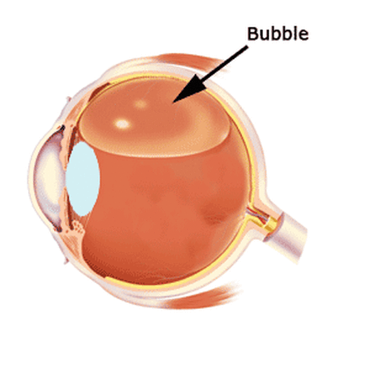
A subset of retinal detachments can be treated by injecting a gas bubble in the eye with a specific head position being necessary after surgery for several days to a week to ensure the retina remains attached and treating the retinal tear with either laser or cryopexy (freezing). This is the only way that a retinal detachment can be treated without a trip to the operating room. Some patients are ideal candidates for this straightforard procedure and can be successfully treated this way avoiding a trip to the OR.
Vitrectomy
Vitrectomy, often referred to as Pars Plana Vitrectomy (PPV), has been used to treat retinal detachments for several decades. More recently, it has become the most commonly chosen surgical option for repairing retinal detachments including when pneumatic retinopexy does not work. By making tiny incisions into the eyeball, instruments are able to remove all the vitreous and subretinal fluid and reattach the retina. The retinal tear or tears that caused the detachment are then treated with laser to cause a permanent adhesive scar in this area and prevent a future detachment. A gas bubble, or less frequently an oil bubble, is instilled in the eye at the end of surgery to maintain the retina in contact with the eye wall as the laser scar matures. Usually a specific head position may be necessary after surgery to ensure the retina remains attached. PPV is performed as a same-day surgery at an outpatient surgery center. Removal of the vitreous may result in progressive clouding of the natural lens in the eye. This is called “cataract” and may necessitate removal of the cloudy lens. In complex cases, a vitrectomy may be combined with a scleral buckle.
Scleral Buckle
Scleral Buckle is the oldest surgical procedure in correcting retinal detachments. A small piece of silicone may be sutured on or around the eye in a fashion that indents the eyeball and brings the retinal break that caused the detachment again in contact with it. This allows the subretinal fluid to reabsorb and the retina to reattach. Sometimes an air or gas bubble is injected at the time of surgery to aid reattachment of the retina. One major advantage of this time-honored technique is that postoperative positioning is not necessary and that no vitreous is removed, preventing premature formation of a cataract (clouding of the natural lens in the eye, which eventually necessitates cataract surgery). This same-day surgery can normally be performed using local anesthesia and allows most patients to resume most daily activities within a few days